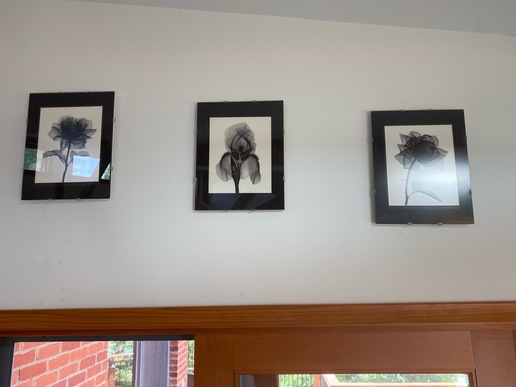
In our continuing frenzy to hang stuff on our walls, we decided those flower x-rays sitting in our storage room needed to see the light of day. There was a big expanse of white in our bedroom over the door to the deck just begging for art. We’d bought the pictures from the artist in 1990, whom we went to visit in his Ann Arbor Hills home. Retired from the dental school, he was a radiologist and avid gardener who had the notion the flowers he was growing might show us something new if he x-rayed them. Indeed they did.
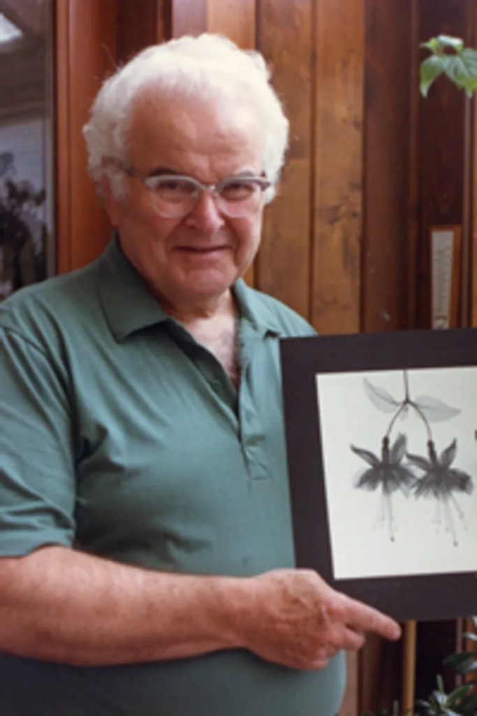
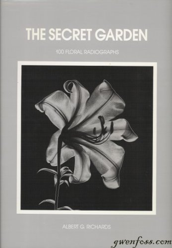
As with many curiosities in this town, it was a piece in the University Record that got me to seek him out. I won’t say he was old, but my late mother-in-law Ruth, who graduated U of M as a dental hygienist in 1953, had him as a professor. He was a kindly host, appreciating the attention, telling us how he developed his technique, still marvelling over what he’d found, enthusiastic as when he made his discovery. He’d just published a book of his radiographs, which may be what prompted the Record piece that led me to him. That book is still in print, available on Amazon (1). It’s nice sized 12 X 9 ½ coffee table book.
I must say, the pics look amazing on our wall. This shot doesn’t do justice to the detail those x-rays show. It’s probably a loss they’re so high, as you really need to get your nose into the image to appreciate fully all the detail.
Professor Richards died in 2008. Born in 1917, he was a year older than my dad. There are some marvelous tributes to him and his work on the web (2,3,4) . A number of his radiographs were in 3-D (4), or even “colorized”. The technique for the latter was so painstaking, he made only a few, and refused to part with one despite Kathy’s imploring. There’s a named award in his honor, the Albert G. Richards Graduate Student Research Grant is designed to assist postgraduate students in conducting applied research in Oral and Maxillofacial Radiology, conferred by AAOMR, the American Academy of Oral and Maxillofacial Radiology (5). U of M published a nice historical vignette (6). His works are displayed in the Getty Museum (7). You can buy his works on Pinterest (8).
Prints of A.G. Edwards’ x-rays are still pretty reasonable on Pinterest. So I’m not going to cash in by owning the works of a deceased artist, it seems. Heck, I don’t even remember how much I paid him for what I’ve got. But the joy Kathy and I will feel as we arise each morning and see those flowers is priceless, and we’ll be forever thanking Professor Richards for that.
References
1. Richards A.G. The secret garden, 100 Floral Radiographs. Almer Co, 1990. https://www.amazon.com/Secret-Garden-100-Floral-Radiographs/dp/096287910X
2. Richards Radiographs. https://flowerxrays.com/bio
3. Contributions from the Museum of Jurassic Technology. The Floral Steroradiographs of Albert G. Richards. https://www.mjt.org/exhibits/alRichards/richards2.html
4. Welcome to the world of Professor ALBERT RICHARDS’ 3-D flower X-rays. https://archivesusie3d.wixsite.com/3-dlegends/albert-g-richards
5. AAOMR. American Academy of Oral and Maxillofacial Radiology. Albert G. Richards Award. https://aaomr.memberclicks.net/albert-g-richards-award
6. The Secret Garden – History. http://www-personal.umich.edu/~agrxray/history.html
7. Getty Museum Colllection. Albert G. Richards. https://www.getty.edu/art/collection/person/104MYZ
8. The Floral Radiographs of Albert G. Richards. https://www.pinterest.com/pin/113715959312162042/
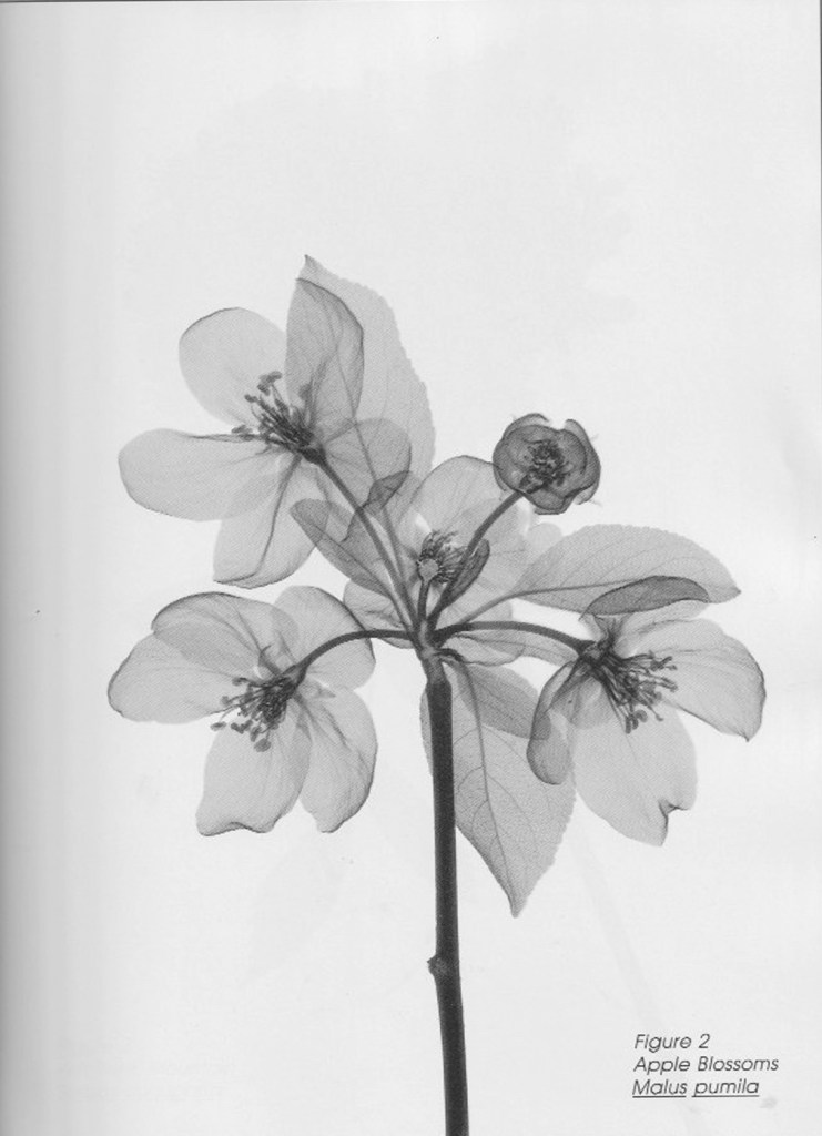
Addendum. When I posted this blog a week and a day ago, I told you about Professor Richards and his technique, but did not do a very good job of showing the results of that work. The pic on the cover of his book (1) and the shot of my own three treasures of his hanging on my bedroom wall hint at the wonder of his works, but hardly capture that wonder. Several of the references have well-detailed, if small, representations (2,3,4,8). Scans from his book aren’t optimal either, but that’s what I did. Here, then, are the first 3 of the 100 radiographs in his book, which appear in alphabetical order, from amaryllis to zinnia. The 9 ½ X 12 book doesn’t quite fit on my 8 ½ X 11 7/8 scanner, and all radiographs are full page, but each has a small border, so my scans did capture each whole picture.
First, an amaryllis (Hippeaeastrum puniceum)

Next, an apple blossom (Malus pumila)
Finally, a blossom from a mountain ash tree (Sorbus aucuparia)

The flower on the cover of The Secret Garden is a lily while mine are 2 roses either side of an iris. Professor Edwards is holding 2 blossoms of a fuschia.
I’ve given you plenty of references to explore about Professor Richards and his work, should you be interested in seeing more or maybe even having your own copy of The Secret Garden for your coffee table. It’ll be like nothing else that rests there.

Interesting…R
LikeLike